Advantages of using BlueShaper files on a maxillary premolar with three canals
Dr. Roberto Estevez shows us the advantages of working with BlueShaper files on a female patient’s maxillary first premolar with three canals, a clinical case that is rarely seen since it only happens in 2.3% of the population (Abella et al – J Endod 2015).
In recent years, many nickel-titanium (NiTi) instruments have been brought to market for canal preparation during endodontic treatments. This has led to an increased use of rotary files by general dentists, because these sixth-generation or newly-emerged instruments have enormous advantages over the first ones that appeared on the market in the nineties.
One of the great advantages lies in the thermomechanical process that occurs during the manufacturing of the instruments, which allows for the creation of rotary files that are more resistant to cyclic fatigue (Shen et al. J Endod 2013).
Recently, in the year 2020, the BlueShaper instruments (Zarc4endo, Gijon-Spain) emerged, which are characterized by their two alloys: pink and blue. This double alloy allows for the combination of the main characteristics of these materials, giving torsional resistance to the first instrument (Z1), and great flexibility and shape memory control to the other instruments (Z2-Z3-Z4).
In the following article, Dr. Roberto Estevez, dedicated exclusively to endodontics at Endodoncia Madrid, shows us an interesting clinical case of a maxillary first premolar with three roots and three canals. This case was managed thanks to the help of CJ-Optik’s Flexion Microscope magnification and the use of BlueShaper files with memory control.
Case Report: Instrumentation of a 3-canal maxillary first premolar with BlueShaper files
A middle-aged patient visited the exclusively endodontic consultation due to pain in the first quadrant. The patient was able to locate the pain by pointing to tooth 14. Upon testing, the patient had negative cold sensitivity on tooth 14, positive percussion and palpation, and physiological probing. Therefore, the diagnosis was pulp necrosis with symptomatic apical periodontitis.
Diagnosis
An out of the ordinary external and internal anatomy could be seen on the diagnostic X-ray (Fig. 1) as there appeared to be three independent roots.
According to research, using the article by Abella et al. (J Endod 2015) as a reference, the incidence of having 3 roots in a maxillary first premolar of a female patient is 2.3%, with two vestibular canals (mesiobuccal and distobuccal) and a palatal canal. Unlike a maxillary molar, it is important to know that the pulp chamber is narrower in the mesiodistal direction, making location and instrumentation of the canal system difficult.
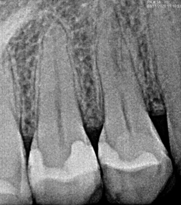
Fig. 1: Diagnostic X-ray showing the complexity of the canal system present in the right maxillary first premolar.
Treatment
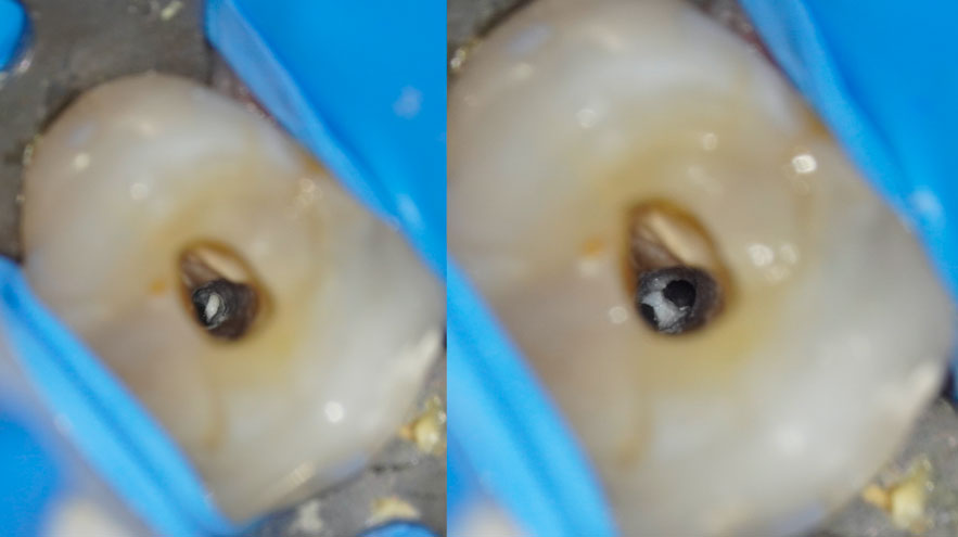
After anesthesia and absolute isolation, an oval opening was made with a round diamond bur, locating two canals (palatal and distobuccal). With the help of magnification and ultrasound the third canal (mesiobuccal) was found, which was very close to the distobuccal one (Fig. 2 and 3).

Fig. 2 and 3: The entrance of the mesiobuccal canal is shown before and after locating and pre-widening with the Z1 file of the BlueShaper system.
The conductometry of the three canals was performed (MB and DB: 19.5 mm and P: 21mm) (Fig. 4 and 5). It is important to note the curvature of the mesiobuccal canal, which can make instrumentation of the canal system difficult.
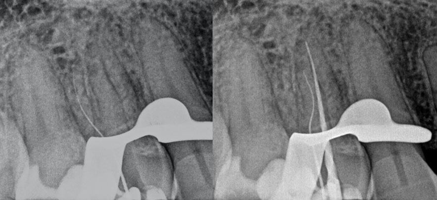
Fig. 4 and 5: The image on the left shows the length of the distobuccal canal. The image on the right shows the longest palatal canal from a mesial view and a double curve present in the mesiobuccal canal.
During all the instrumentation process, irrigation was being done with 4.2% sodium hypochlorite using a beveled irrigation needle 27g. Due to the proximity of the mesio and distobuccal canals, the decision was made to insert the first rotary file of the BlueShaper system (Z1:14/02) manually (Fig. 6), and once verified, to place the E-Connect S endodontic motor (Eighteeth) on the head of the file and operate it, to know at all times which canal was being worked on.
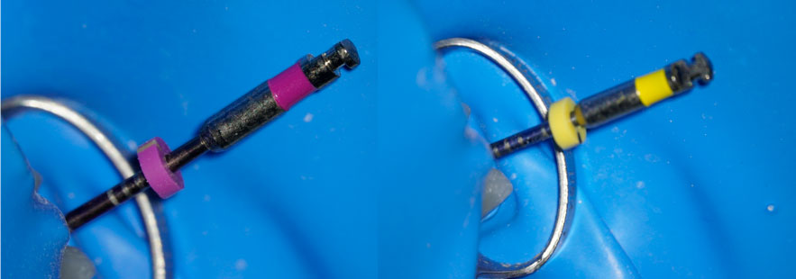
Fig. 6: Z1 and Z3 files of the BlueShaper system inserted manually prior to their instrumentation with the Eighteeth motor.
This is one of the great advantages of BlueShaper. Due to their shape memory control, instruments can be pre-curved manually, the curved shape is maintained and therefore adapts to the selected canal. Once widened at the entrance, preparation is easier; always maintaining constant irrigation and patency with our manual file 10 allows for a safe preparation.
Afterward, and as indicated in the following sequence (Fig. 7), Z2 and Z3 instruments were inserted into all the canals. In the palatal canal the Z4 file (06/25) was also used, while stopping at the Z3 instruments in the vestibular canals (06/20).
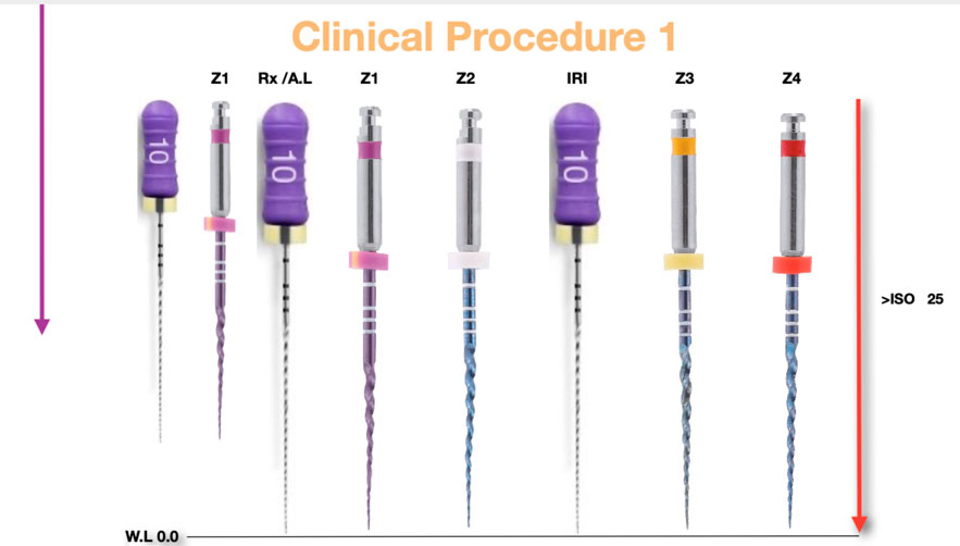
Fig. 7: Recommended instrumentation sequence for canal system instrumentation with BlueShaper files.
After instrumentation, a final irrigation protocol was performed based on sonic activation of the irrigants: sodium hypochlorite, 17% liquid EDTA, and again sodium hypochlorite. Finally, the canals were dried and their obturation was performed.
Obturation
The obturation was carried out using the vertical condensation technique with the Fast Pack device by Eighteeth (tip 40/03). Using the microscope allowed for the obturation to be controlled at all times, especially as canals were so close in this case (Fig. 8 and 9). The backfill or filling of the coronal two-thirds was carried out with the injection device by the same manufacturer (Eighteeth) using a 23g needle and medium consistency gutta-percha.
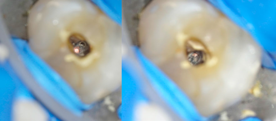
Fig. 8 and 9: images showing the downpack or obturation of the apical third of the distobuccal and mesiobuccal canals.
The post-obturation X-rays (Fig. 10 and 11) show the adaptation of the gutta-percha three-dimensionally, sealing a lateral canal present in the palatal canal, and adequately respecting the original anatomy of the canal system present in this right maxillary first premolar.
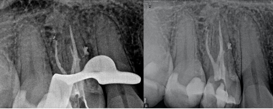
Fig. 10 and 11: Condensation and immediate postoperative X-rays where the three-dimensional adaptation of the gutta-percha to the canal system can be observed.
Finally, the provisional material was placed, an analgesic and/or anti-inflammatory prescribed in case of discomfort, and patient was referred to their general dentist for final restoration.

Dr. Roberto Estévez
Doctor in Dentistry and Master in Endodontics (Universidad Europea of Madrid). Coordinator of the Master of Endodontics Program at the Universidad Europea of Madrid. Lecturer for endodontic courses and conferences at a national and international level. Author of various articles in national and international scientific journals. Exclusively practicing endodontics since 2004.
More information
If you liked this clinical case reporting treatment of a maxillary premolar with three canals, please let us know. Get in touch with us, we are happy to help you.
Write us an email at hola@zarc4endo.com.


